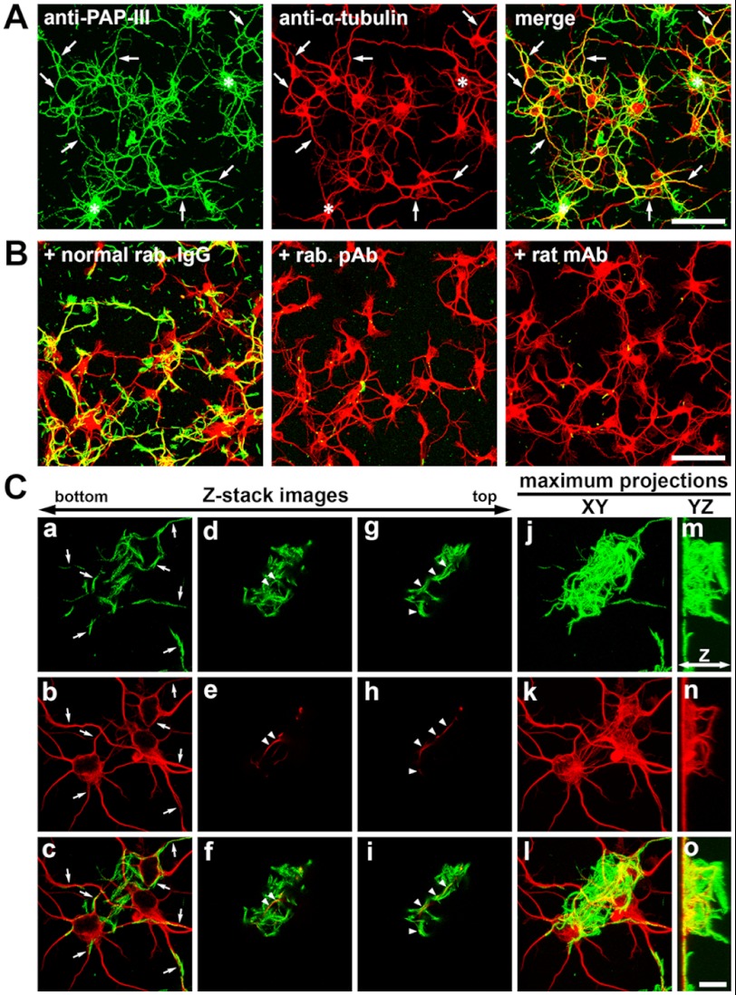
FIGURE 5.
Association of exogenously added ΔN-PAP-III fibers with neurons. ΔN-PAP-III fibers produced in 25 mm Tris (pH 7.5) containing 150 mm NaCl were added to the culture media of primary cortical neurons precultured for 12–18 h. Twenty-four hours after incubation, the preparations were fixed and immunostained with anti-PAP-III (green) and anti-α-tubulin (red) antibodies. A, ΔN-PAP-III fibers associated with neurites (arrows) as well as somata. Complexes of accumulated fibrils were often formed and accumulated on somata (asterisks). B, the associations were blocked by preincubating ΔN-PAP-III fibers with rabbit polyclonal (+rab. pAb) or rat monoclonal anti-PAP-III (+rat mAb) antibody, but not with normal rabbit IgG (+normal rab. IgG). C, high magnification Z-stack images of regions where large complexes of ΔN-PAP-III fibers accumulates on somata (indicated by asterisks in A). Superficial images of the coverslips show a clear association of ΔN-PAP-III fibers with neurites (a–c, arrows). The top images show the upward growth of some neurites into accumulated fibrillar complex (d–f and g–i, arrowheads). Maximum projections of XY (j–l) and YZ (m–o) planes. Neurites grow upward only in the presence of fibrillar complexes (m–o). Scale bars, 50 μm (A and B) and 10 μm (C).

