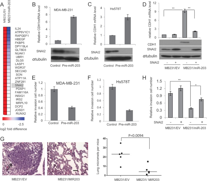
FIGURE 2.
miR-203 targets SNAI2 and inhibits breast cancer metastasis. A, heat map showing 25 predicted targets that were suppressed by miR-203 through microarray analysis. B, transfection of pre-miR-203 in MDA-MB-231 cells inhibited SNAI2 and induced CDH1 as determined by Western blot and real time RT-PCR, respectively. C, transfection of pre-miR-203 in Hs578T cells inhibited SNAI2 and induced CDH1 as determined by Western blot and real time RT-PCR, respectively. D, miR-203 up-regulated CDH1 by targeting SNAI2. Both MB231/miR-203 cells and MB231/EV cells were transduced with retroviruses expressing SNAI2 without the 3′-UTR or control vector. The expression of SNAI2 was examined by Western blot, and the expression of CDH1 was examined by Western blot and real time RT-PCR. *, p < 0.05; **, p < 0.01. E and F, transfection of pre-miR-203 inhibited breast cancer invasion in both MDA-MD-231 and Hs578T cells as determined by Matrigel invasion assays. The data are mean ± S.D. of two independent experiments preformed in triplicates. G, the restoration of miR-203 in MDA-MB-231 cells inhibited lung metastasis in nude mice. Metastatic tumors were examined by H&E staining. Arrow, metastatic tumor tissues. The graph shows the quantification of the total number of nodules in individual lungs (n = 5). Student's t test was used for the significance calculation. Scale bar, 50 μm. H, the restoration of SNAI2 partially rescued miR-203-mediated inhibition. Error bars represent S.D. micromets, microscopic metastases.

