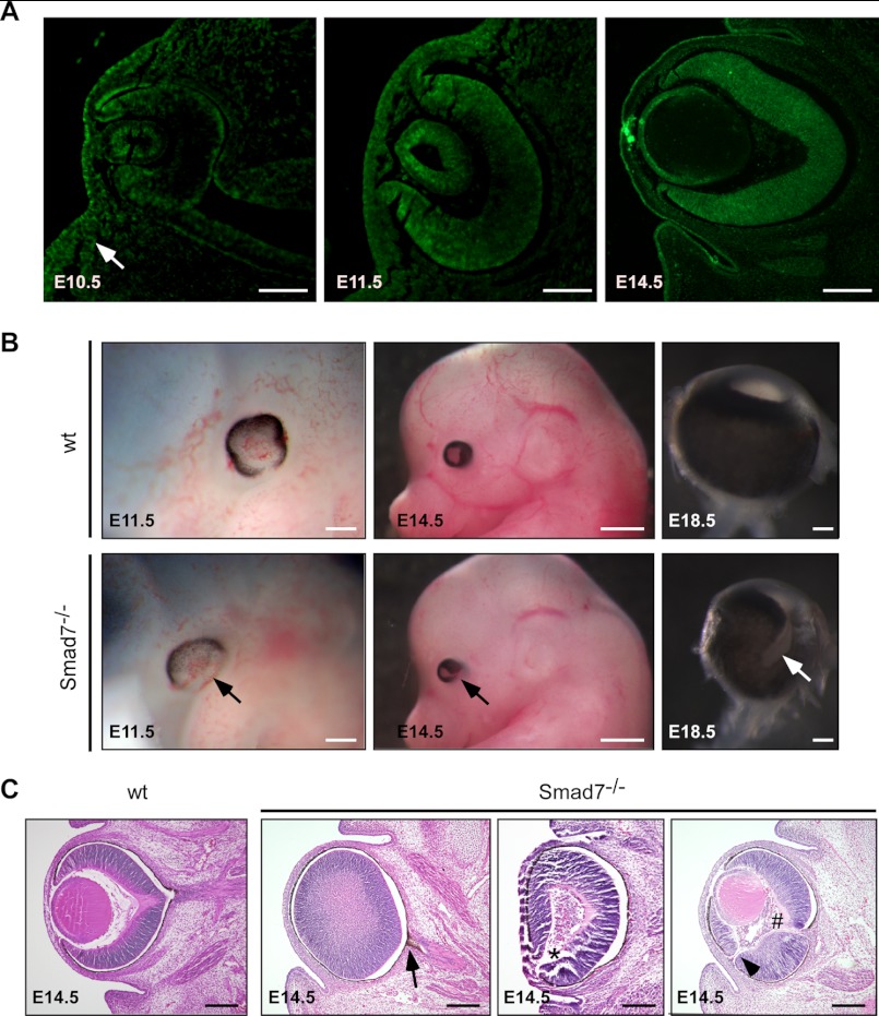
FIGURE 1.
Deletion of Smad7 leads to ocular defects. A, Smad7 protein was examined by immunohistochemistry in the developing eyes of mouse embryos at E10.5, E11.5, and E14.5 using the frontal sections. In addition to the lens and retina, Smad7 protein was detected in the surrounding mesenchyme of the eye (indicated by an arrow) at E10.5. B, whole mount view of ocular phenotype in Smad7 null embryos. Pigmentation is deficient in the ventral retina at E11.5 compared with the control (marked by arrow). Coloboma and microphthalmia are observed at E14.5 and at E18.5 in Smad7-deficient embryos (marked by arrows). Only eyeballs are shown for E18.5. C, transverse section stained with H&E shows pigmentation in the optic stalk (marked by arrow), absence of the lens (marked by asterisk), hyperplastic primary vitreous (marked by # sign), and discontinuity of the retina (marked by arrowhead) in Smad7-deficient embryos. Scale bars: 50 μm (A, first and second panels); 100 μm (A, third panel; B, first and third panels; C, all images); 400 μm (B, second panels).

