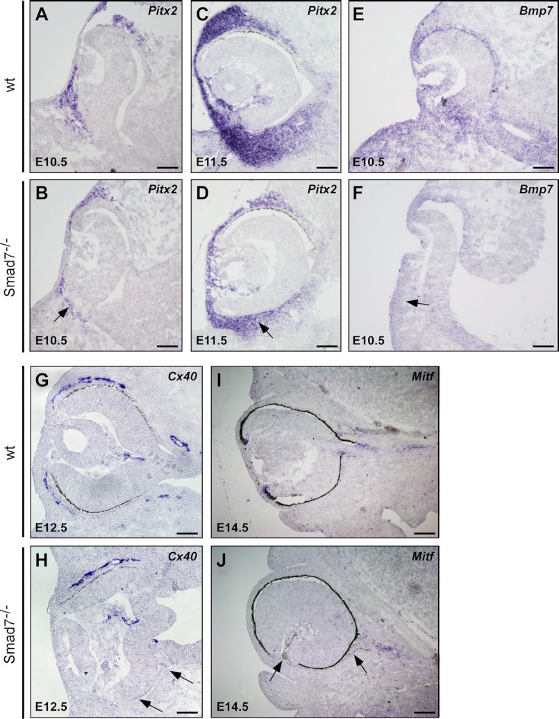
FIGURE 4.
Altered gene expressions in the periocular mesenchyme of Smad7 null mice. The eyes of the control and Smad7-deficient embryos were used in in situ hybridization to analyze the expression of genes in the periocular mesenchyme including Pitx2 (A–D), Bmp7 (E and F), Cx40 (G and H), and Mitf (I and J) at different stages of the embryo. Note that the eyes of Smad7 mutant embryos have reduced expression of Pitx2 in the optic cup (marked by an arrow), decreasing BMP7 level in the surrounding mesenchyme (indicated by an arrow), defective blood vessel (Cx40-positive region) surrounding the eye (marked by an arrow), and ectopic Mitf expression (marked by an arrow). Scale bars: 50 μm (A–H) and 100 μm (I and J).

