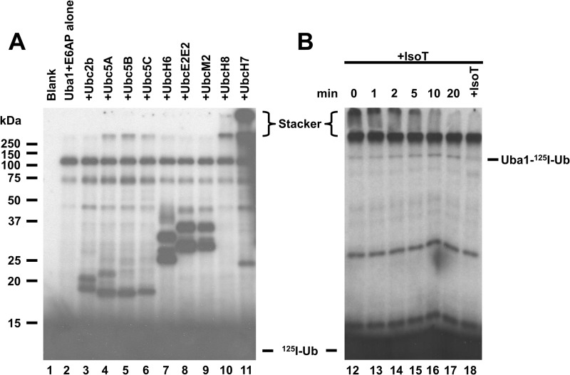
FIGURE 1.
UbcH7 is the cognate E2 for E6AP. A, 125I-ubiquitin conjugation assays containing 16 nm GST-E6AP were conducted for 10 min in the absence (lane 2) or presence of 100 nm concentrations of the indicated E2 (lanes 3–11) then quenched with SDS sample buffer and resolved by 12% (w/v) SDS-PAGE under reducing conditions as described under “Materials and Methods.” The resulting gel was dried, and 125I-ubiquitin conjugates were visualized by autoradiography. B, incubation identical to that of lane 11 was depleted of ATP by the addition of 6 IU apyrase (0 min) then recombinant isopeptidase T (+IsoT) was added to a final concentration of 17 nm. Aliquots were taken at the indicated times and quenched with SDS sample buffer. Additional isopeptidase T was added at 20 min to a final concentration of 34 nm, then the reaction was allowed to proceed for an additional 20 min before being quenched with SDS sample buffer. Samples were resolved and visualized as in panel A. Mobility of molecular weight markers are shown to the left. Positions of the monoubiquitinated activating enzyme (Uba1-125I-Ub) and free ubiquitin (125I-Ub) are as indicated. The position of the 5% (w/v) stacker is shown by brackets.

