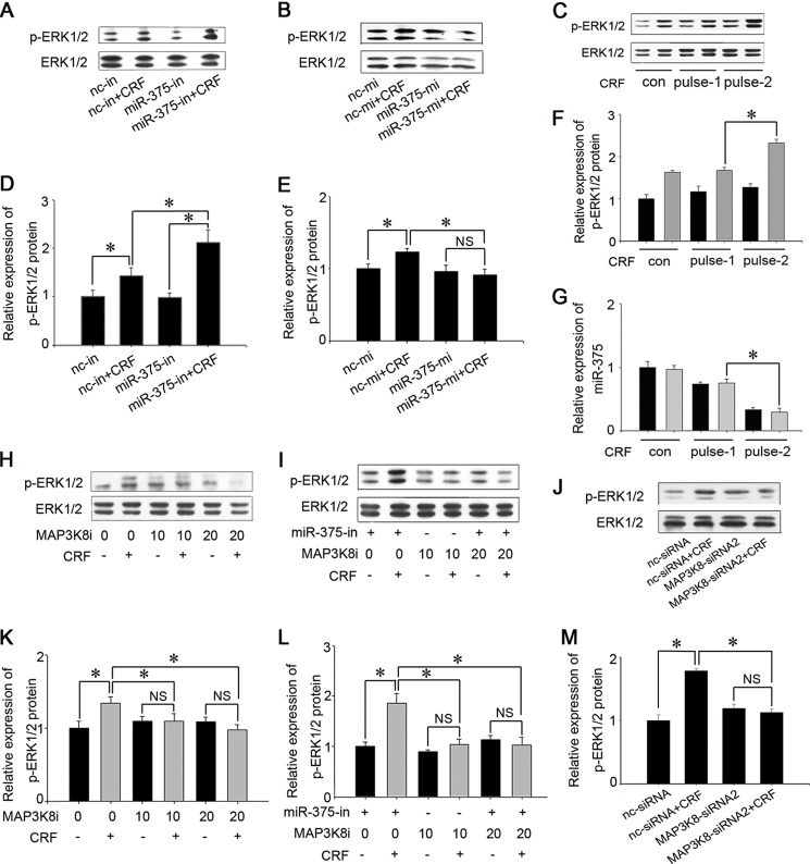
FIGURE 6.
miR-375 and MAP3K8 regulate ERK1/2 phosphorylation. A, analysis of ERK1/2 and P-ERK1/2 proteins. AtT-20 cells were transfected with miR-375 inhibitors and cultured with 100 nm CRF for 10 min, and protein expression levels were analyzed by Western blotting. B, analysis of ERK and P-ERK1/2 proteins. AtT-20 cells were transfected with miR-375-mi for 24 h and 100 nm CRF for 10 min. C, effect of CRF (100 nm) plus stimulation on ERK1/2 phosphorylation. G, effect of CRF (100 nm) pulse stimulation on miR-375 expression. H, analysis of ERK and p-ERK1/2 proteins. AtT-20 cells were treated with MAP3K8i. CRF was added 1 h later and cultured for 10 min. I, analysis of ERK and p-ERK1/2 proteins. AtT-20 cells were transfected with miR-375-in for 24 h, and then 20 μm MAP3K8i was added for 1 h. The cells were then treated with 100 nm CRF for 10 min. J, analysis of ERK and p-ERK1/2 proteins. AtT-20 cells were transfected with MAP3K8 siRNA2 for 24 h and then treated with 100 nm CRF for 10 min. D–F and K–M, quantification of p-ERK1/2 protein levels. Data are presented as means ± S.E. (n = 3). *, p < 0.05; NS, not significant (p > 0.05) (ANOVA). con, control; nc-mi, nc mimic; nc-in, nc inhibitor.

