
FIGURE 1.
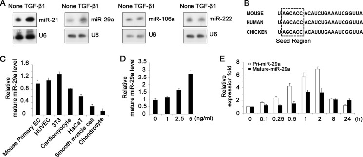
TGF-β1 up-regulates miR-29a expression in ECs. A, Northern blot analysis of miRNAs in HUVECs treated with or without 5 ng/ml TGF-β1, showing up-regulation of miR-21 and miR-29a by TGF-β1. B, sequences of mature miR-29a in mouse, human, and chicken. C, real-time PCR analysis of mature miR-29a expression in several kinds of primary cells and cell lines, including mouse primary ECs, human primary ECs (HUVEC), cardiomyocytes, smooth muscle cells, chondrocytes, a fibroblast cell line (NIH-3T3), and a human keratinocyte line (HaCaT). D, real-time PCR analysis of mature miR-29a expression in bEnd.3 cells. TGF-β1 increased miR-29a expression in a dose-dependent manner (1–5 ng/ml). E, real-time PCR analysis of primary and mature miR-29a expression in response to 5 ng/ml TGF-β1 at different time points.