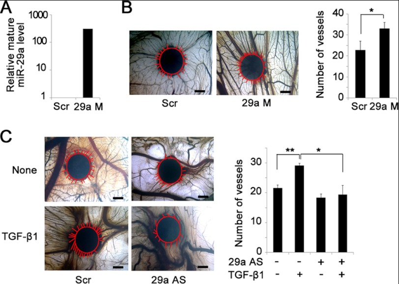
FIGURE 3.
MiR-29a mediates TGF-β1-induced angiogenesis. A, real-time PCR analysis confirmed miR-29a overexpression with miR-29a mimic transfection (29a M) compared with scrambled sequence transfection (Scr). B, miR-29a mimic promoted new vessel growth in a CAM assay (left panel). Newly formed blood vessels were quantified (right panel). Scale bar = 1 mm. *, p < 0.05. n = 6. Red lines indicate the edge of the filter paper and the newly formed vessels around the filter paper. C, inhibition of miR-29a blocked TGF-β1-induced angiogenesis in the CAM. Filters soaked with TGF-β1 alone, with miR-29a antisense oligonucleotides (29a AS), or with the scrambled sequence were applied onto the CAM (left panel). Newly formed blood vessels were quantified (right panel). Scale bar = 1 mm. *, p < 0.05; **, p < 0.01. n = 6. The red line indicates the vessels around the filter paper.

