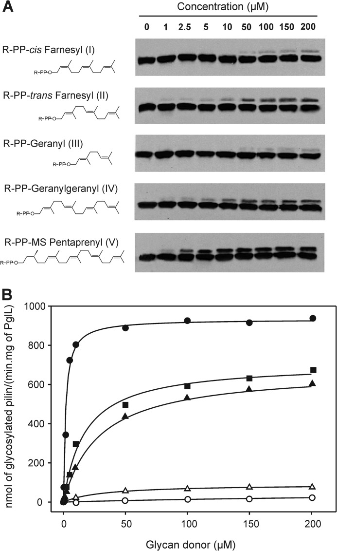
FIGURE 3.
Activity of PglL with glycan donor substrates carrying the same glycan portion (E. coli O86 O antigen) but polyisoprenoid chains varying in length and geometry. A, structures of the substrates and Western blot analysis of the resultant in vitro glycosylation assays. The observed signals correspond to glycosylated and non-glycosylated pilin (upper and lower bands, respectively; antibody anti-pilin SM1). R-PP denotes the sugar pyrophosphate attached to each lipid. B, activity plots and kinetic adjustments for all the glycan donors. Black circles, substrate V; black squares, substrate IV; black triangles, substrate II; white triangles, substrate I; and white circles, substrate III.

