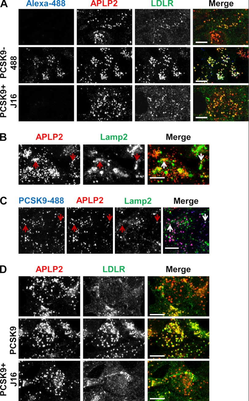
FIGURE 7.
Characterization of the endocytic route of APLP2 and colocalization with PCSK9 and LDLR. A, APLP2 (red) and LDLR (green) surface staining on HepG2 cells in the absence (top), or presence of PCSK9–488 (middle, blue), or presence of PCSK9/J16 (bottom). B, colocalization of internalized anti-APLP2 monoclonal antibody (red) with Lamp2 (green). Examples are indicated by arrows. C, colocalization of internalized anti-APLP2 monoclonal antibody (red) with internalized PCSK9-488 (blue) and with Lamp2 (green). Examples are indicated by arrows. D, APLP2 (red) colocalization with LDLR (green) with or without exogenously added PCSK9 or PCSK9/J16 as indicated. Scale bars, 10 μm.

