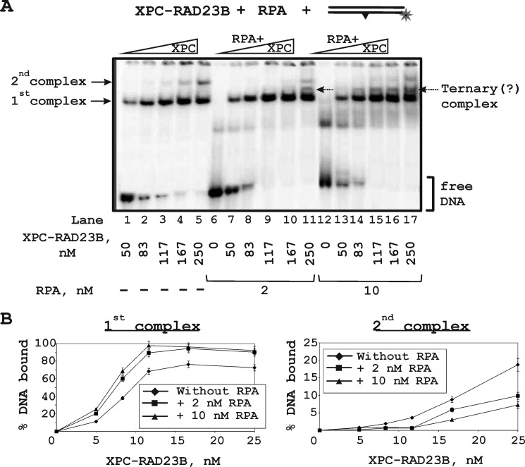
FIGURE 7.
Binding of the XPC-RAD23B to damaged DNA duplex in the presence of different RPA concentrations. A, the damaged DNA duplex was incubated at a concentration 10 nm with the indicated concentrations of XPC-RAD23B without/with various RPA concentrations: 2 or 10 nm. At the concentrations analyzed, the XPC-RAD23B formed two protein-DNA complexes with different electrophoretic mobility. B, the quantitative analysis of the protein-DNA complexes (from A). Percentages of bound DNA were plotted against XPC-RAD23B concentrations.

