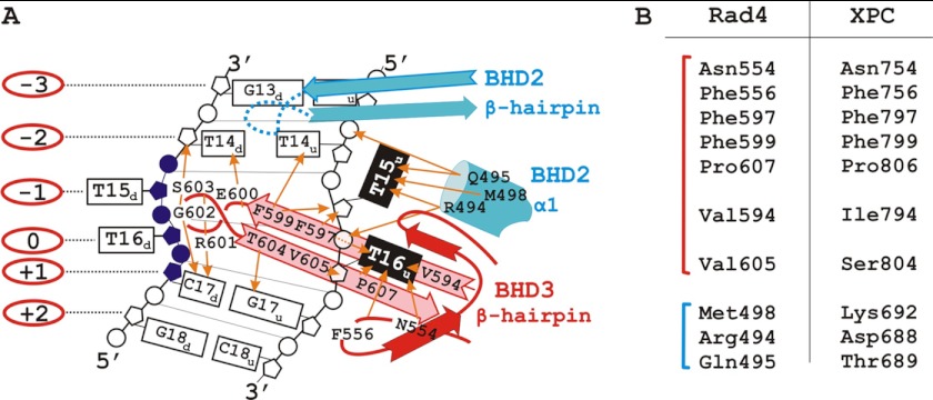
FIGURE 8.
A, schematic representation of the interactions of the BHD2-BHD3 domains with a 4-bp damaged DNA segment according to the Rad4-Rad23 crystal structure data (17). The BHD2 and BHD3 domains are marked in blue and red, respectively. The flipped-out thymidines of the undamaged strand (T15u and T16u) are in black; the disordered, cyclobutane pyrimidine dimer-linked thymidines (T15d and T16d) and the damaged strand segment are in purple; numbers in the red ovals indicate numbering of nucleotides in the damaged strand relative to the T16d nucleotide; the protein side chain contacts to DNA are marked with orange arrows. B, the BHD2 and BHD3 residues (blue and red, respectively) that contact the damaged DNA segment and the corresponding residues from the XPC structure (17). Analogous and identical residues in these positions suggest that human and yeast orthologs bind to DNA in the similar way.

