
Fig 3.
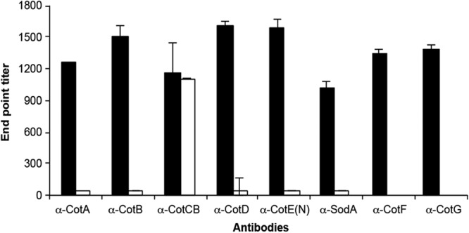
Surface location determined by “whole-spore ELISA.” Microtiter plates were coated with spores, which were probed with antibodies (α) to each of the spore coat proteins. Spores were either wild-type 630Δerm (black bar) or the corresponding isogenic Clostron mutant (unfilled bar), with the exception of anti-CotF and anti-CotG, where mutants were not available. Naive serum was used also for comparison, and basal levels were subtracted.