
Fig 4.
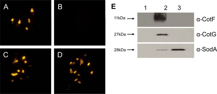
CotF, CotG, and SodA. Based on bioinformatics analysis, three additional spore coat proteins were identified: CotF (tyrosine rich), CotG (putative catalase), and SodA (superoxide dismutase). Polyclonal antibodies to each of these recombinant proteins were used to examine surface location on the 630Δerm spore coat. Panels A to D show immunofluorescent labeling of spores: 630 spores labeled with anti-SodA antibodies (A), spores of the sodA::CT394s mutant labeled with anti-SodA (B), 630 spores labeled with anti-CotF (C), or 630 spores labeled with anti-CotG (D). Spores showed no labeling with preimmune serum (data not shown). Panel E shows Western blots probed with anti-CotF, anti-CotG, and anti-SodA. Lane 1, extracts of proteins found in the supernatant of vegetative cells that had been sonicated (10 bursts of 30 s with cooling); lane 2, extracts from the supernatant of pelleted spores that had been sonicated (10 bursts of 30 s with cooling); lane 3, proteins extracted from spores using a protein extraction buffer (sodium borate-SDS-DTT) described previously (18). Every lane carried 10 μg of protein, determined using the Bradford assay.