
Fig 1.
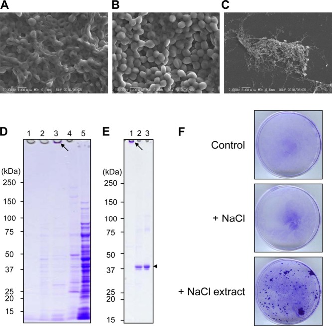
Isolation of biofilm matrix proteins and cell wall-anchored proteins from S. aureus biofilms. The surface structures of biofilms produced by S. aureus SH1000 without (A) and with (B) 1 M NaCl treatment were observed by scanning electron microscopy. (C) The structure of the NaCl-extracted biofilm matrices from the SH1000 biofilm was also analyzed. (D) The culture supernatant fraction (lane 1), 10 mM Tris-HCl (pH 8.0) washing fraction (lane 2), 1 M NaCl-extracted fraction (lane 3), cell wall fraction obtained by lysostaphin treatment (lane 4), and cytoplasmic fraction yielded after sonication (lane 5) were dissolved by SDS-PAGE and stained with CBB. An arrow indicates insoluble materials stacked on the well. (E) A 1 M NaCl extract from the SH1000 biofilm was treated without (lane 1) or with (lane 2) 0.2 mg/ml dispersin B and then subjected to SDS-PAGE. Dispersin B was also subjected to SDS-PAGE as a control (lane 3). The arrow indicates insoluble materials stacked on the well, and the arrowhead represents dispersin B. In panels D and E, molecular sizes are given at the left. (F) The effect of 1 M NaCl extracts from biofilms on cell aggregation and attachment to an abiotic surface was examined. S. aureus MS4, which forms less biofilm, was incubated at 37°C for 1 h in the absence or presence of a 1 M NaCl extract from SH1000 biofilms. Cells attached to polystyrene dishes were stained with crystal violet. NaCl solution (1 M) instead of 1 M NaCl extract was added as a negative control.