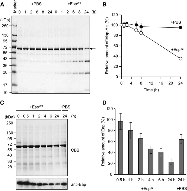
Fig 4.
Esp-dependent degradation of Eap in vitro and in S. aureus biofilm. (A) Purified recombinant Eap (1 μM) was incubated in the presence of EspWT at 37°C for 24 h. As a control, PBS was added instead of EspWT. (B) Band intensities on the gel (A) were estimated using the ImageQuant system. Data points represent the means and standard deviations of results from three independent experiments. The standard deviation is less than the size of the symbol if no error bars are seen. (C) The time-dependent degradation of Eap by EspWT in the biofilm was analyzed by SDS-PAGE with CBB staining and Western blotting using an anti-Eap polyclonal antibody. As a control, PBS was added instead of Esp. Molecular sizes are given at the left. (D) Band intensities on the X-ray film (C) were estimated using ImageQuant. The means and standard deviations of triplicate determinations are represented.

