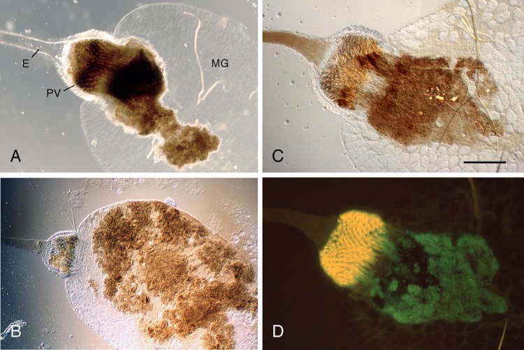
Fig 2.
Fragile biofilm produced by PhoP− Y. pestis in the flea gut. Digestive tracts of X. cheopis fleas infected with Y. pestis KIM6+ (A), KIM6+ ΔphoP (B), or KIM6+ ΔphoP (pGFP) (C and D) were dissected and examined by light (A to C) and fluorescent (D) microscopy. The proventriculus (PV) of the flea (A) is filled and blocked with a dense cohesive biofilm that extends into the midgut (MG). The biofilm produced by the phoP mutant is less cohesive and is usually confined to the MG (B) or attached only peripherally to the posterior ends of the autofluorescent spines of the PV (C and D). The examples shown are representative of several flea dissections. Bar = 0.1 mm. E, esophagus.

