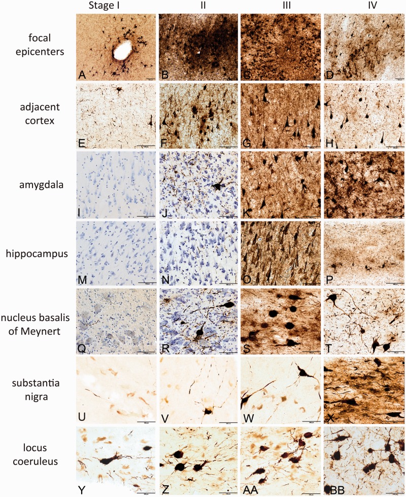
Figure 4.
Hyperphosphorylated tau pathology in the four stages of CTE. In stage I CTE (first column), p-tau pathology is found in limited discrete perivascular foci (A), typically at the depths of sulci or around small vessels. There is mild p-tau pathology in cerebral cortices neighbouring the epicentres (E). There is no or minimal p-tau pathology in the amygdala (I) or CA1 of hippocampus (M). Occasional p-tau neurites are found in the nucleus basalis of Meynert (Q) and substantia nigra (U); isolated neurofibrillary tangles are present in the locus coeruleus (Y) in stage I. In stage II CTE (second column), there is spread of pathology from focal epicentres (B) to the superficial layers of adjacent cortex (F). The medial temporal lobe shows only mild neurofibrillary pathology, including amygdala (J) and CA1 hippocampus (N). Nucleus basalis of Meynert (R) and locus coeruleus (Z) demonstrate moderate p-tau pathology as neurofibrillary tangles and neurites; the substantia nigra (V) shows only modest pathology. In stage III, p-tau pathology is severe and widespread throughout the frontal, insular, temporal and parietal cortices. The cortical epicentres and depths of the sulci often consist of confluent masses of neurofibrillary tangles and astrocytic tangles (C). The intervening cortices show advanced neurofibrillary degeneration (G). The amygdala (K), hippocampus (O) and entorhinal cortex demonstrate marked neurofibrillary pathology. The nucleus basalis of Meynert shows dense neurofibrillary tangles (S); the locus coeruleus (AA) shows advanced neurofibrillary pathology, and the substantia nigra is moderately affected (W) in stage III CTE. In stage IV CTE, there is widespread p-tau pathology affecting most regions of the cerebral cortex and medial temporal lobe with relative sparing of the calcarine cortex. Astrocytic tangles are prominent, and there is marked neuronal loss in the cortex, amygdala and hippocampus. Phosphorylated-tau neurofibrillary tangles are reduced in size and density. The cortical epicentres show severe neuronal loss and prominent astrocytic tangles (D); similar changes are found throughout the frontal, temporal and parietal cortices (H). The amygdala demonstrates intense gliosis and p-tau neuronal and glial degeneration (L). The hippocampus is sclerotic with marked neuronal loss, gliosis, ghost neurofibrillary tangles and astrocytic tangles (P). The nucleus basalis of Meynert shows marked neurofibrillary pathology and gliosis (T); the substantia nigra (X) and locus coeruleus (BB) show advanced neurofibrillary pathology. All images: CP-13 immunostained 50 -µm tissue sections, some counterstained with cresyl violet, all scale bars = 100 µm.

