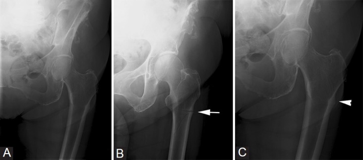
Figure 1 (A-C).

Patient 1: The initial frontal radiograph of the left hip (A) was reported as normal. The patient presented again after 1 month, when a frontal radiograph (B) showed an incomplete fracture (arrow) involving the lateral aspect of the proximal femoral shaft, below the level of the greater trochanter. A review of the initial radiograph (C) showed subtle breaking of the lateral cortex (arrowhead)
