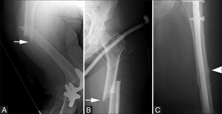
Figure 2 (A-C).

Patient 2: Radiograph of the right femur (A) shows the typical complete fracture of the proximal femur (arrow). Anteroposterior radiograph of the left hip (B) of the same patient 5 months later shows a similar fracture (arrow). Post-operative radiograph after surgical fixation (C) shows beaking and thickening of the lateral cortex at site of the original fracture (arrowhead)
