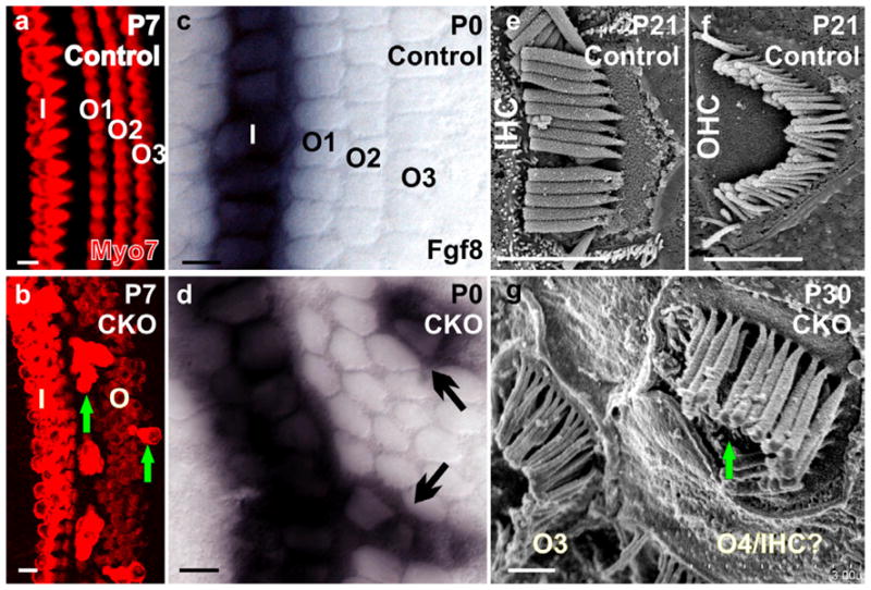
Figure 4. Absence of Neurod1 leads to ectopic IHCs formation in the OHC region.

Myo7a immunocytochemistry reveals one row of IHCs and 3 rows of OHCs in the postnatal day (P) 7 control mice (a). Neurod1 conditional null (CKO) mice show 2 rows of IHCs with multiple rows of OHCs as well as some extensively stained Myo7a positive ectopic IHCs in the position of OHCs (b, arrows in b). Fgf8 is expressed only in the IHCs in control mice (c) but is expressed in some OHCs (arrows in d) in Neurod1 CKO mice. Scanning electron microscopy confirms that ectopic IHCs have the thick stereocilia typical of IHCs but in the configuration of OHC stereocilia (e–g, arrow in g). I, IHCs; O1-O3, OHC row 1–3. Modified after (Jahan et al., 2010a). Bar indicates 100 μm except e–g, where it indicates 1 μm.
