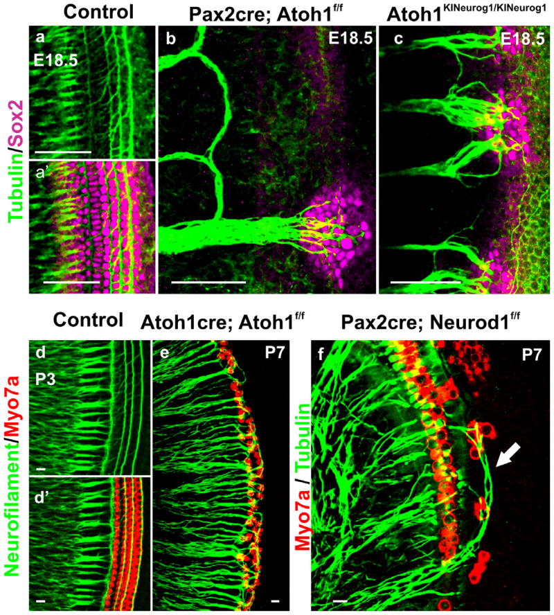
Figure 6. HC defects affect differentially the innervation pattern.
Tubulin and Sox2 immunochemistry at E18.5 control mice show the projection of type I fibers to IHCs and the type II to OHCs (a, a′). Conditional deletion of Atoh1 with Pax2-cre results in the absence of HCs differentiation with atypical looping fibers modiolar to the OC, except for some fibers reaching to the Sox2 positive patches of undifferentiated OC cells (b). Homozygotic Neurog1 misexpression in Atoh1 knockin mice (Atoh1KINeurog1/KINeurog1) results in increased density of innervation as well as projection of radial fibers to Sox2 positive OC cells (c) indicating that Neurog1 can partially compensate for the lack of Atoh1. Neurofilament and Myo7a co-labeling displays type I and type II fiber projection to the IHCs and OHCs, respectively in the P3 control mice (d, d′). Delayed deletion of Atoh1 using Atoh1-cre reveals formation of only two rows of OHCs, resulting in aberration of innervation (e): all fibers, possibly including type I fibers, project to the remaining OHCs in the absence of their target cell, the IHCs, in this mutant (e). Neurod1 loss results in ectopic formation of IHCs in the region of OHCs, in addition to the formation of multiple rows of IHCs and OHCs (f). Tubulin co-labeling with Myo7a demonstrates the atypical overshooting of the type I fibers to the ectopic IHCs in the region of OHCs as well as disorganization of overall fiber projection (f). These mutants indicate that the variable alterations in the OC HCs can differentially modify the type-specific innervation pattern. Modified after (Jahan et al., 2010b; Jahan et al., 2012; Pan et al., 2012b). Bar indicates 100 μm in a–c and 10 μm in d–f.

