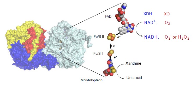
Fig. (1).

Crystal Structure of Bovine XOR. Left; Homodimer structure of bovine XOR. The N-terminal (in red), the C-terminal (in blue) and the intermediate (in yellow) domains contain the iron-sulfur centers, the molybdopterin and the FAD centers. Right: Cofactor arrangements of the enzyme. Figures were generated from PDB ID 1F4Q. Arrows show the directions of electron flow during catalysis. The reduced FAD reacts with either NAD+ or oxygen to produce NADH or hydrogen peroxide (H2O2) or superoxide (O2-). FADH2 reacts with O2 to produce H2O2 , while FADH produces O2- [61, 63]. (The color version of the figure is available in the electronic copy of the article).
