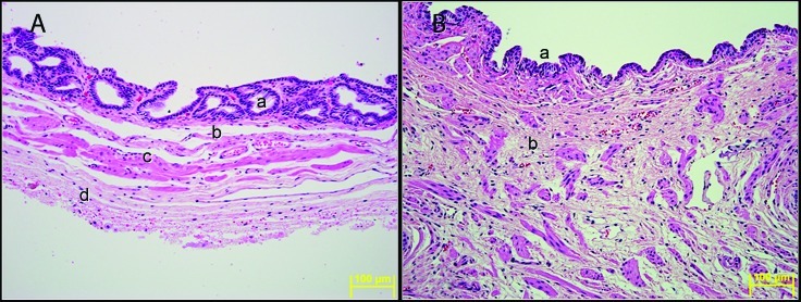
Figure 5.
(A) Vesicular gland. a, mucosa with glands; b, submucosa; c, tunica muscularis; d, adventitia. (B) Vesicular gland near the ejaculator duct. a, mucosal epithelium; b, thick wall of loose connective tissue with bundles of smooth muscle fibers. Hematoxylin and eosin stain. Magnification, 200×.

