
Figure 1.
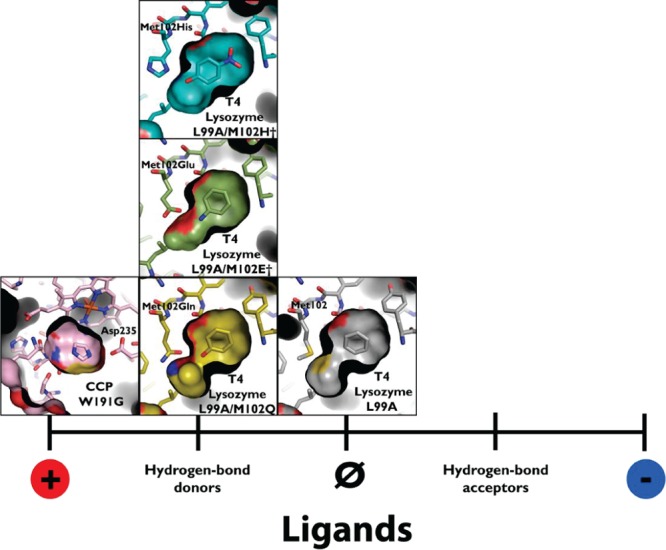
Model cavities to test molecular docking and protein–ligand interactions. These cavities range from the L99A cavity in T4 lysozyme (represented by PDB 181L(28)), which binds exclusively apolar, hydrophobic ligands, to the W191G cavity in cytochrome C peroxidase (CCP) (represented by PDB 1AES(73)). Intermediate to these are the more polar T4 lysozyme cavities L99A/M102Q (PDB 1LI2), which adds a polar glutamine residue to the cavity and L99A/M102E† (PDB 3GUN(55)), which adds a neutral glutamic acid, and L99A/M102H† (PDB 4EKQ(37)), the focus of this study, which adds a histidine residue to the cavity. These intermediate cavities bind both nonpolar and polar, hydrogen-bond donating ligands.