
Figure 10.
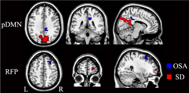
Brain regions with significantly differences in rsFC of the pDMN and RFP between the patients with OSA (blue) and the sleep-deprived subjects (red). L, left; OSA, obstructive sleep apnea; pDMN, posterior default-mode network; R, right; RFP, right frontoparietal network; rsFC, resting-state functional connectivity; SD, sleep deprivation.