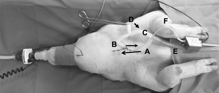
Figure 1.
Breathing circuits in the anesthetized pig. A, rostral tracheal tube; B, caudal tracheal tube; C, tube connecting rostral and caudal trachea; D, tube to atmosphere for tracheal breathing in the open state; E, tube to negative pressure device; F, thin tube for the registration of sublaryngeal pressure that was advanced into the rostral tracheal tube. Arrows on tubes A and B show the direction of the tubes in the trachea. In the setting illustrated in the figure the pig is in a situation of nasal breathing, with the clamp closing the tube to atmosphere. Removing the clamp from position D and putting it onto the connecting tube between rostral and caudal trachael tubes (C, arrow) leads to tracheal breathing, a situation in which actuation of the negative pressure device for the collapsibility test directs the negative pressure to the upper airway in an inspiratory direction via tube E.

