
Figure 3. NA excites GCs in the MOB and AOB by activating α1A-ARs.
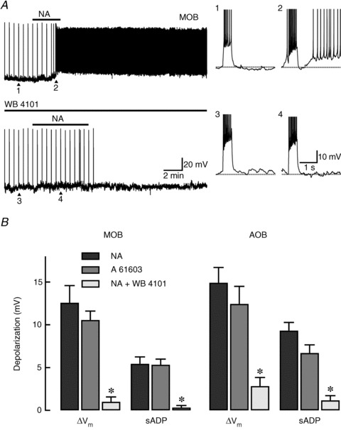
A, top trace: recording from a GC in the MOB where bath application of NA (10 μm) depolarized the cell and produced an sADP that resulted in a series of spontaneous action potentials. In this cell, spike trains, which appear compressed as vertical lines in the time axis, were elicited every 30 s by a depolarizing current stimulus (30 pA, 500 ms). The right traces correspond to those indicated by the arrowheads and are shown in an expanded time scale. In these traces, the dotted lines indicate the pre-stimulus membrane potential. 1: under control conditions, the current stimulus produced seven spikes that were followed by a small afterhyperpolarization (ΔVm: 1 mV). 2: post-application of NA, the same stimulus elicited 10 spikes and an sADP (7 mV) that greatly enhanced the cell's excitability. Bottom trace: in the same cell, application of a low concentration of the α1A-AR selective antagonist WB 4101 (30 nm) completely abolished the depolarization and sADP produced by NA. 3 and 4: expanded responses to the current pulse at the points indicated with arrowheads (control and NA in the presence of WB 4101, the resting membrane potential is −62 mV). B, summary of the α1A-AR-mediated changes in excitability of GCs in the MOB and AOB. The α1A-AR selective antagonist WB 4101 (30 nm, white bars) greatly reduced the NA-induced depolarization (ΔVm, black bars) in the MOB and AOB (n= 12 respectively). Likewise, application of WB 4101 significantly decreased the sADP in both regions (*P < 0.0006; MOB: n= 9; AOB: n= 6). Similarly, the NA-induced depolarization and sADP were mimicked by the α1A-AR selective agonist A 61603 (1 μm, grey bars) in the MOB and AOB (n= 9, respectively). AOB, accessory olfactory bulb; AR, adrenergic receptor; GC, granule cells; MOB, main olfactory bulb; NA, noradrenaline; sADP, slow afterdepolarization.