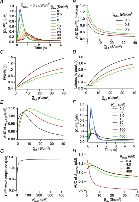
Figure 3. A-type K+ channels regulated calcium release from ER stores during calcium waves in a three-cylinder neuronal model.

A, effect of the A-type conductance in the oblique dendrite on [Ca2+]c during a Ca2+ wave. [Ca2+]c traces were recorded at a distance of 8 μm from the branch point with different densities of the A conductance in the oblique dendrite. B–E, area under the curve (AUC) for the [Ca2+]c transients depicted in A (B), full width at half maximum, FWHM (C) and latency-to-peak for these [Ca2+]c transients (D) and AUC for the flux of Ca2+ through InsP3 receptors,  (E), in achieving these transients, plotted as functions of A-conductance density,
  . Plots are shown for different densities of the L-type Ca2+ channel,
  . F and G, time-dependent [Ca2+]c changes (F) and the amplitude (G) of Ca2+ wave, computed in the presence of mobile buffers at different affinities (Kmob) to Ca2+ binding. Ca2+ wave was initiated with 100 μm mobile buffer with the same protocol as in A. H, AUC for
  plotted as functions of A-conductance density,
  , when the Ca2+ wave was initiated in the presence of 100 μm mobile buffer and with different values of Kmob.
