
Figure 2. ATP- and Glu-evoked Ca2+ signals in neurones and astrocytes in a rat hippocampal neurone–astrocyte co-culture.
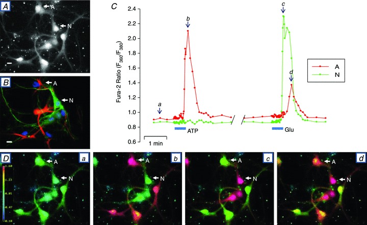
A, fura-2 stained neurones and astrocytes (F360 excitation) immediately before initiating the Ca2+ signalling experiment. Cell A is an astrocyte and N is a neurone. B, immunostained cells from the same field as in A. Immediately after the Ca2+ signalling experiment, the cells on the coverslip were fixed and cross-reacted with anti-NF-H antibodies to identify neurons (N, green) and with anti-GFAP antibodies to identify astrocytes (A, red). All nuclei were stained with DAPI. C, time course of changes in the fura-2 F360/F380 ratio, illustrating the effects of 0.5 μm ATP and 4 μm l-glutamate (Glu). D, fura-2 F360/F380 ratio images captured at time points a–d indicated in C. These data are a representative example of 4 similar, fully analysed experiments.