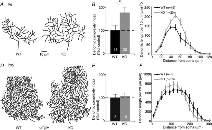
Figure 1. Increased morphological complexity of Purkinje cells in 5-HT3A receptor knockout mice at postnatal day (P) 9 but not at P35.

A, reconstructed Purkinje cells from wild-type (WT) and 5-HT3A receptor knockout (KO) mice at P9. B, dendritic complexity index of Purkinje cells at P9 indicates an increased dendritic complexity in 5-HT3A receptor knockout mice in comparison to WT mice. C, Sholl analysis indicates an increased dendritic length, specifically at 30–60 μm from the soma, in 5-HT3A receptor knockout mice at P9. D, reconstructed Purkinje cells from WT and 5-HT3A receptor knockout mice at P35. E, the dendritic complexity index of WT and 5-HT3A receptor knockout mice does not reveal any difference at P35. F, Sholl analysis does not show any topological difference between Purkinje cells from WT and 5-HT3A receptor knockout mice at P35.
