
Figure 5. MIM-S1 inhibits MIM-mediated endocytosis and membrane protrusions.
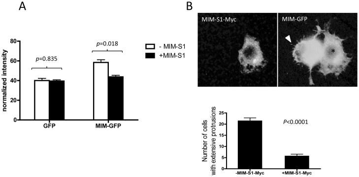
(A) Cells co-expressing MIM-S1-Myc and MIM-GFP or cells expressing GFP only were treated with Bio-Tfn for 5 min. Uptake of Bio-Tfn was analyzed as described in Experimental. While Tfn uptake was markedly increased in cells expressing MIM-GFP, no significant increase was observed in cells expressing both MIM-GFP and MIM-S1-Myc. (B) 293T cells were transiently transfected with MIM-GFP and MIM-S1-Myc, fixed and stained with anti-Myc antibodies. The stained cells were inspected by immunofluorescent microscopy. Original magnification, 600×. A cell showing expression of MIM-GFP only exhibited many filopodia-like long membrane protrusions as indicated by an arrow head. A cell, as indicated by *, expressing both MIM-GFP and MIM-S1-Myc failed to display these MIM characteristic protrusions. Quantification (lower panel) showed that cells co-expressing MIM-GFP and MIM-S1-Myc had about 70% less MIM-mediated membrane protrusions than those expressing MIM-GFP alone.