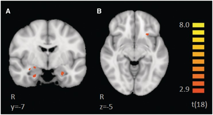
Fig. 6.
Results of PPI analysis, using a left medial prefrontal cortex seed (functionally defined in the slow > fast contrast) and an anatomically defined bilateral amygdala and insula mask. The figure illustrates coactivation in the left mPFC, bilateral amygdala and left insula during CT-targeted affective, slow touch relative to fast touch, which does not target CT-afferents.

