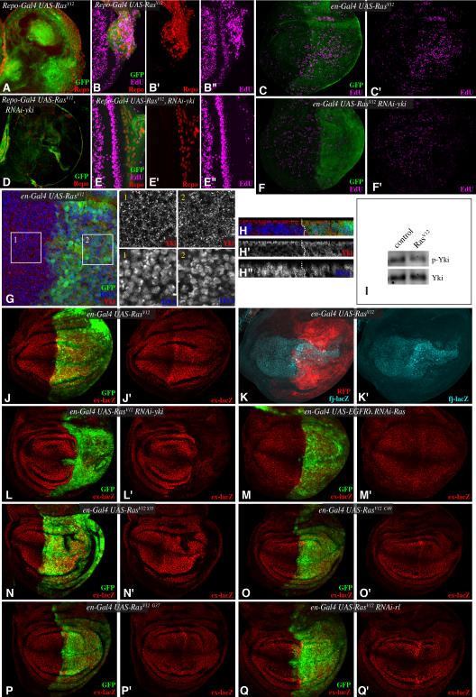
Figure 3. EGFR regulates Yki through a Ras-Raf-MAPK pathway.
Third instar brain lobes (A,D), eye discs (B,E) or wing discs (C,F) stained for Repo (red) or EdU (magenta) from larvae expressing (A,B,D,E) repo-Gal4 UAS-mCD8:GFP (green) or (C,F) en-Gal4 UAS-GFP (green) and A-C) UAS RasV12, or D-F) UAS RasV12 UAS-Yki-RNAi. G,H) Wing disc stained for Yki (red) and DNA (Hoechst, blue) from en-Gal4 UAS-GFP (green) UAS-RasV12. Higher magnifications of single channels of Yki and DNA stains from boxed regions 1 and 2 are shown at right, H shows vertical sections through the disc. I) Western blot on lysates of wing discs expressing RasV12 (UAS-RasV12 tub-Gal4 tub-Gal80ts, induced for 3 days at 29 °C), probed with anti-Yki and anti-phospho168 Yki antibodies. (J, L-Q) Third instar wing discs, stained for β-gal expressed by ex-lacZ (red) from larvae expressing en-Gal4 UAS-GFP (green) and J) UAS-RasV12, L) UAS-RasV12 UAS-Yki-RNAi, M) UAS-EGRλ UAS-Ras-RNAi, N) UAS-RasV12S35, O) UAS-RasV12C40, P) UAS-RasV12G37, Q) UAS-RasV12 UAS-rl-RNAi. K) Wing discs, stained for β-gal expressed by fj-lacZ (cyan) from larvae expressing en-Gal4 UAS-mRFP (membrane bound RFP, red) and UAS-RasV12. See also Fig. S2.

