Figure 4.
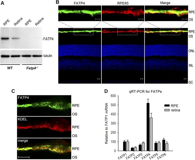
FATP4 is the predominant FATP in RPE. A, Immunoblot analysis of FATP4 and tubulin in retinas and RPE from 129S2/Sv (WT) and Fatp4−/− mice. B, Immunohistochemistry showing FATP4 in the WT mouse RPE and its colocalization with RPE65. RPE, outer segments (OS) of photoreceptors, outer nuclear layer (ONL), inner nuclear layer (INL), and ganglion cell (GC) layer are indicated. Scale bars, 10 μm. C, Immunohistochemistry showing colocalization of FATP4 with KDEL, an ER marker antibody, in the mouse RPE. Scale bar, 10 μm. D, qRT-PCR showing relative expression levels of six FATP family members in the mouse RPE and retina. Relative FATP mRNA levels are shown as the percentage of FATP1 mRNA level. Error bars indicate SD (n = 3).

