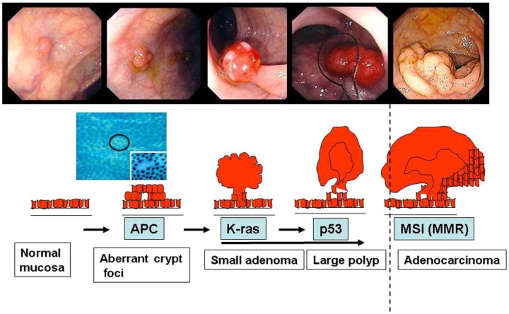
Figure 4.
Colorectal carcinogenesis from normal to invasive cancer.
The slides on the top are from colonoscopy after cleaning preparation. The genes involved in each step are also indicated. High magnification shows aberrant crypt foci is needed to detect precancerous lesions in macroscopic normal mucosa. APC, adenomatous polyposis coli; K-ras, Kirsten rat sarcoma; MMR, mismatch repair; MSI, microsatellite instability.

