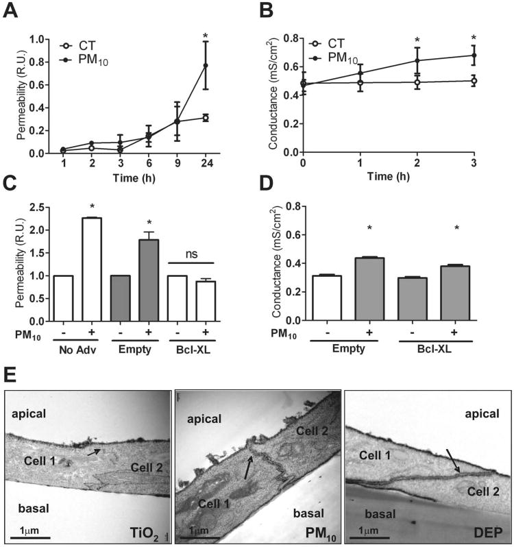
Figure 1. PM disrupts alveolar epithelial barrier.
Panel A & B. Primary rat AEC were plated on inserts and at day 3 of isolation were treated with PM10 (20μg/cm2). Permeability to 4 kDa FITC-dextran (A) and Gt (B) were measured at different time points (1, 2, 3, 6, 9 & 24 hours). Panel C & D. 24 hours after infection of AEC with adenovirus containing Bcl-xL plasmid, cells were treated with PM10 (20μg/cm2) and permeability to FITC-dextran was measured at 24h (C) and Gt was determined at 3h after treatment with PM10 (20μg/cm2) (D). Panel E. Primary rat AEC were treated for 3h with 50μg/cm2 of TiO2, PM10 or DEP, they were then fixed and stained with La3+ on the apical side of the insert and observed under transmission electron microscopy. Electron dense La3+ penetrates the TJ (arrows) into the intercellular space after treatment with PM10 and DEP but not with TiO2. Graph represents mean ± SEM (n=3). * p<0.05 when compared with control.

