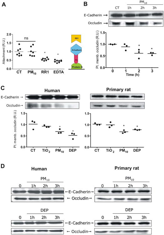
Figure 2. PM reduces occludin abundance at the plasma membrane in AEC.
Panel A. Human AEC were treated with calcein-AM and allowed to attach to a protein G plate coated with E-cadherin-Fc chimera (schematic representation on the right). Cells were treated with PM10 (20μg/cm2) for 3h; wells then were washed and fluorescence of remaining cells was measured. Panel B. Human AEC were treated with PM10 (20μg/cm2) for 1, 2 and 3 hours, surface proteins were then labeled with biotin and pulled down with streptavidin beads, WB of these proteins is shown. Panel C. Human and primary rat AEC were treated with 20μg/cm2 of TiO2, PM10 or DEP and surface protein were labeled with biotin and pulled down with streptavidin beads and analyzed by WB. Panel D. Total cell lysate of human and rat AEC were analyzed by WB after treatment with PM10 and DEP (50μg/cm2). Representative blots and quantitative analysis of three independent experiments are shown. Graph represents mean with dots representing each individual experiment. * p<0.05 when compared with control.

