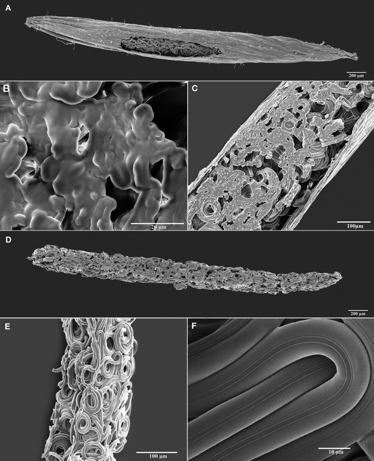
Fig. 4.
Scanning electron micrographs (SEM) of juveniles of Anguina agrostis (Steinbuch, 1799) Filipjev, 1936 dissected from seed galls of Agrostis stolonifera L. A: Seed gall sliced longitudinally to expose the juveniles contained within. B: Cross-section revealing juveniles tightly packed within the seed gall showing the body contents that appear gel-like in the SEM and oozing out of the cut nematodes and coalesce with each other. C: Close-up of a seed gall sliced longitudinally to exposes the anhydrobiotic nematodes and the contents from their bodies. D: Mass of juvenile nematodes dissected from a seed gall in an anhydrobiotic state. E: Close-up showing coiled anhydrobiotic juveniles dissected from the seed gall. F: Close-up of juveniles revealing that the morphology of the body wall remains smooth, undistorted, and free from any artifact normally associated with the loss of all free water molecules.

