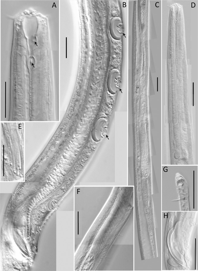
Fig. 2.
Light microscopic pictures of the male of Neotobrilus nicsmolae n. sp. (A) Lip region (arrows at anterior and posterior teeth). (B) Ventromedian supplements. (C) Reproductive system. (D) Pharyngeal region. (E) Flagelloid sperm. (F) Ductus ejaculatoris. (G) Subterminal setae. (H) Spicules. Scale: A, B, G = 20 μm; C–D, F = 40 μm; E = 30 μm; H = 50 μm.

