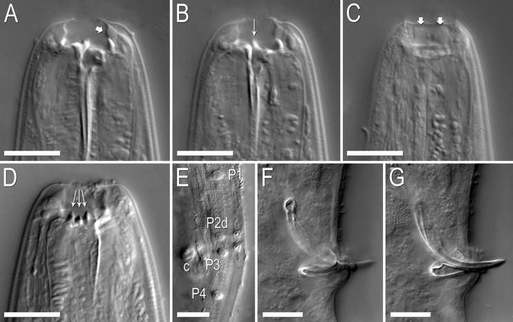
Fig. 4.
Nomarski micrographs of Pristionchus bucculentus n. sp. Scale bars are 10 μm. (A–C) Stoma of a single eurystomatous female in right lateral view through three focal planes. (A) Dorsal plane, showing dorsal tooth and bulging cheilostom (arrow) with thin internal (medial) walls. (B) Right subventral plane, showing right subventral tooth with a narrow, elevated apex. (C) Right subventral plane at right lateral margin of stoma, showing cheilostomatal bulges separated by very weak incision. (D) Left subventral plane of the stoma of a eurystomatous female in left lateral view, showing three regularly shaped, conical denticles. (E) Cuticular surface at male tail in oblique left-ventral view, showing the diagnostic arrangement of genital papillae P1–P4, including with respect to the cloacal opening (c). (F–G) The copulatory organ of a single male individual in right lateral view through two focal planes. Rounded, ventrally skewed manubrium is highlighted in (F). The short, shallow terminal curvature of the gubernaculum is highlighted in (G).

