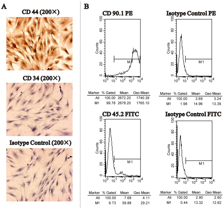
Figure 1. Detection of surface markers of MSCs.
A. Immunocytochemistry results showed that MSCs were positive for CD44 (upper), and negative for CD34 (middle) and isotype control (lower). B. Flow cytometry analysis of MSCs. Almost the entire tested MSCs showed positive for CD90 (left upper), and negative for CD45 (left lower), isotype control Ig G2 PE (right upper) and isotype control Ig G1 FITC (right lower).

