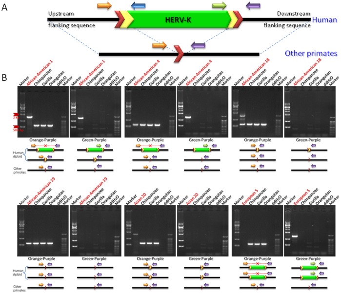
Figure 3. Variable polymorphic patterns of a HERV-K118 in human diploid genomes.
Human-specific HERV-K118 insertion locus was amplified by PCR using the genomic DNAs of human population and other primates as template. (A) A typical primate HERV-K element. The ∼7.5 kb structure of the HERV-K internal region is shown in green. Yellow chevrons are LTRs (∼1 kb) and red chevrons are target site duplications (TSDs). (B) Gel chromatographs show PCR products of targeted human-specific HERV-K loci on a panel containing human three non-human primates. High bands indicate the presence of an insertion, while low bands indicate its absence. Orange and purple arrows indicate primers designed in the conserved flanking regions of all species. Green arrows indicate internal primers designed within the human-specific HERV-K. As shown in the gel pictures, human-specific HERV-K insertion loci exhibit a variety polymorphic patterns in human diploid genomes.

