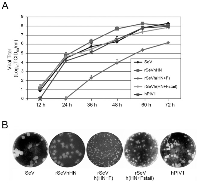
Figure 2. Virus growth kinetics and plaque formation of rSeVs.

(A) Multi-step growth curve of the viruses in LLC-MK2 cells. Cells were infected with wt or chimeric viruses at MOI 0.01 and incubated at 34°C. Aliquots of infected cell supernatants were collected at indicated times after infection and viral titers of supernatants were determined in LLC-MK2 cells. (B) Plaque formation of the wt and rSeVs. LLC-MK2 cells were infected with SeV, rSeVhHN, rSeVh(HN+F), rSeVh(HN+Fstail) or hPIV1 and cultured at 34°C with medium containing agarose. Plaques were identified using crystal violet staining.
