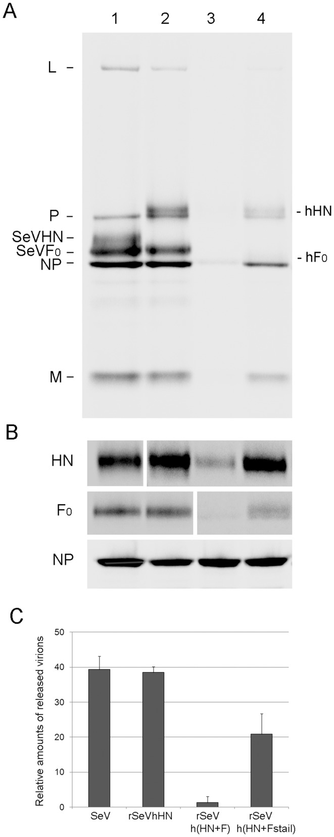
Figure 3. Virus production from infected cells.
(A) Virion production from LLC-MK2 cells infected with SeV (lane 1), rSeVhHN (lane 2), rSeVh(HN+F) (lane 3), or rSeVh(HN+Fstail) (lane 4). Cells were infected at a MOI of 1 and labeled with [35S] Met/Cys for 16 h. Labeled progeny virions released from the cells were purified and analyzed by SDS-PAGE. (B) Viral proteins produced in infected cells. HN, F and NP proteins in [35S]-labeled cell lysates as described in (A) were immunoprecipitated using anti-SeV or hPIV1 HN, F or NP antibodies. (C) Amounts of NP in released virions (A) were quantified and shown as the average of three independent experiments with standard deviations.

