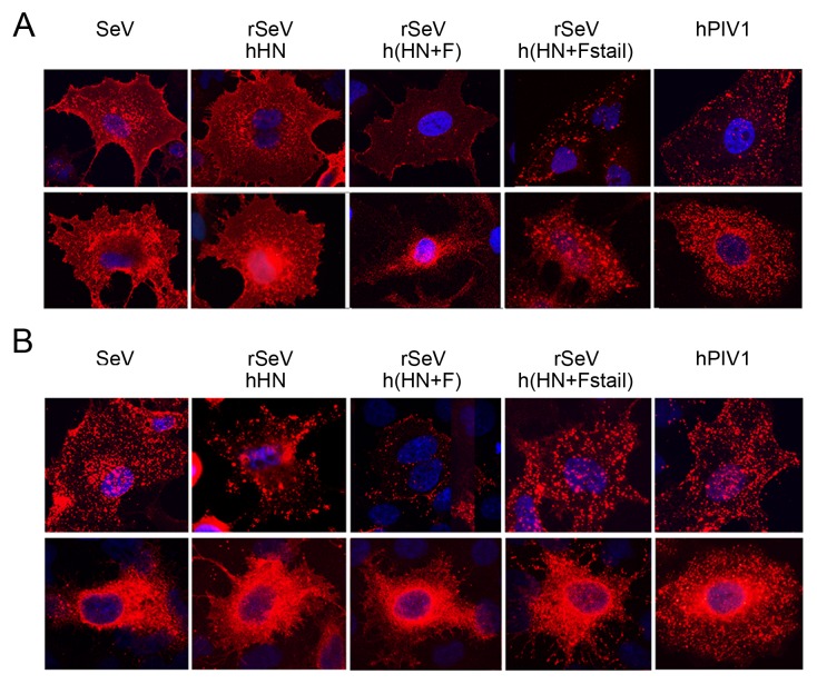
Figure 4. Surface and subcellular localization of HN and F proteins.
A549 cells were infected with SeV, hPIV1 or rSeVs and incubated for 16 h at 34°C. Cells were then fixed and treated with mAbs against SeV or hPIV1 F (A) or HN (B) without (upper panels) or with (lower panels) permeabilization. Anti-mouse IgG-Texas Red was used as secondary.

