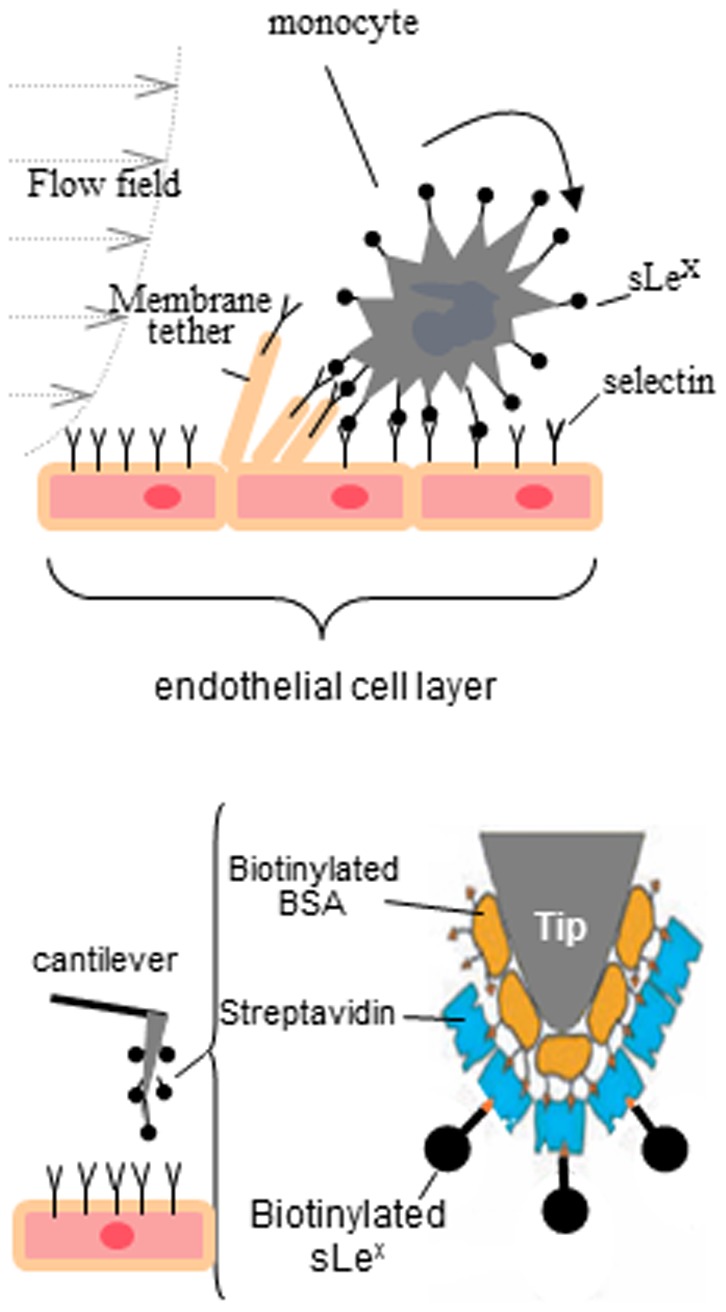
Figure 1. Schematic descriptions of membrane tether formation and biofunctionalization for the AFM cantilever tip.

Membrane tether formation mediated by sLex-selectin bonding during a monocyte rolling on the endothelial layer (upper) and the strategy using AFM cantilever tips bio-functionalized by sLex to characterize the mechanics of membrane tether adhesion (lower). (modified from Yves F. Dufrêne, 2008).
