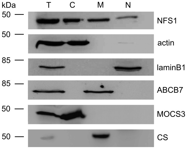
Figure 1. Immunodetection of NFS1 and MOCS3 after subcellular fractionation of HeLa cells.
Total protein extracts (T), cytosol (C), mitochondria (M), and nucleus (N) were prepared separately from 80–90% confluent HeLa cells to avoid cross contaminations of the compartments. Proteins of each cellular fraction were analyzed by immunoblotting using the following antibodies: anti-NFS1 (top panel), anti-γ-actin as cytosolic marker control (second panel), anti-laminB1 as nuclear marker (third panel), anti-ABCB7 as mitochondrial inner membrane marker (fourth panel), anti-MOCS3 as cytosolic marker (fifth panel), and anti-citrate synthase [6] as mitochondrial matrix marker (bottom).

