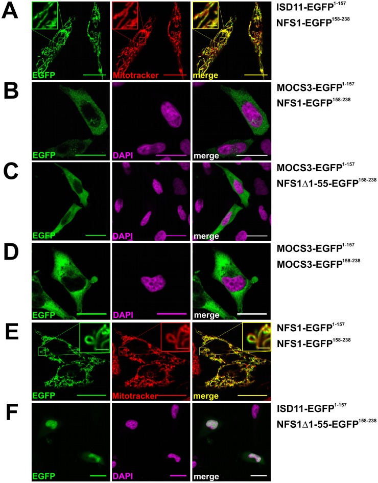
Figure 4. Analysis of NFS1 and MOCS3 interactions by using the split-EGFP system in HeLa cells.
Subcellular EGFP assembly of different split-EGFP fusion proteins was analyzed in HeLa cells by confocal fluorescent microscopy. The following fusion proteins were expressed after cotransfection (assembly of EGFP1–157 and EGFP158–238 resulted in a green pseudocolor): A, ISD11-EGFP1–157 and NFS1-EGFP158–238; B, MOCS3-EGFP1–157 and NFS1-EGFP158–238; C, MOCS3-EGFP1–157 and NFS1Δ1-55-EGFP158–238; D, MOCS3-EGFP1–157 and MOCS3-EGFP158–238; E, NFS1-EGFP1–157 and NFS1-EGFP158–238; F, ISD11-EGFP1–157 and NFS1Δ1-55-EGFP158–238. Mitochondria of HeLa cells were visualized with MitoTracker® DeepRed (red) or the nuclei were visualized with DAPI stain (magenta). Merged pictures are shown right (either resulting in a yellow or white color). Scale bars, 20 µm; scale bars in the insets, 2 µm.

