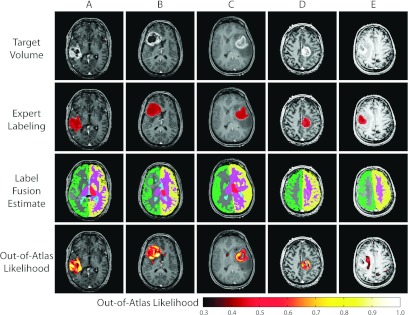
Figure 3.

Qualitative results for the detection of malignant gliomas. Five representative examples are presented. For each example, the target volume, expert labeling, label fusion estimate, and the out-of-atlas likelihood are presented. The first four examples represent cases where the tumor region is correctly identified. The last example represents the outlier case [seen in Fig. 2c] in which the cancerous region was almost completely missed.
